№ 21 · HEALTH
Dental X-rays: What the Radiation Dose Actually Means for Safety
June 06, 2026 · QDRO
Every year, millions of patients shift uncomfortably in the dental chair while a lead apron is draped over their chest, wondering — with varying degrees of anxiety — whether this small rectangle of film is doing something quietly harmful. The concern is understandable. Radiation carries a cultural weight that its actual dosimetry rarely justifies.
The numbers, when you look at them squarely, are genuinely reassuring. But "reassuring" is not the same as "trivial," and the story has enough nuance to be worth understanding properly.
The Dose in Context: Microsieverts and Background Radiation
Radiation dose in medicine is measured in sieverts — a unit that accounts not just for the amount of energy deposited in tissue but for how biologically damaging that particular type of radiation is. For dental imaging, we work in microsieverts (μSv), one-millionth of a sievert.
A standard set of four bitewing X-rays — the most common dental radiograph, used to detect cavities between teeth — delivers approximately 5 μSv of effective dose. A full-mouth series of periapical images comes in at around 35 μSv with rectangular collimation and up to 170 μSv with round collimation, depending on equipment and technique (PMID 18762634).
Compare that to background radiation. Every person on Earth receives ionizing radiation continuously from cosmic rays, naturally occurring radon in buildings, and radioactive isotopes in soil and building materials. The global average is roughly 2.4 millisieverts per year — about 6.6 μSv per day (UNSCEAR 2008 Report, Sources and Effects of Ionizing Radiation). A single bitewing series, in other words, represents less than one day's worth of unavoidable background exposure.
A transatlantic flight from London to New York adds about 50–80 μSv from increased cosmic ray exposure at altitude. A chest CT scan delivers around 7,000 μSv. Mammography: approximately 400 μSv. Dental radiography sits at the extreme low end of the diagnostic imaging spectrum — a fact that is consistent across national regulatory bodies and independent dosimetric studies.
This is not a dismissal of the question. It is a calibration. Risk should be proportionate to actual magnitude, and for routine dental X-rays that magnitude is very small.
CBCT: A Different Conversation
Cone beam computed tomography (CBCT) has become increasingly common in dental practice over the past two decades. It offers three-dimensional imaging of the jaw, teeth, and surrounding bone — invaluable for implant planning, orthodontic assessment, and complex surgical cases. But it comes with meaningfully higher doses than conventional 2D radiography.
Depending on the field of view, resolution settings, and the specific machine, CBCT doses range from approximately 19 μSv to 368 μSv across the 14 scanners measured in the SEDENTEXCT dosimetry study (PMID 21196094) — and older or large-field protocols can push effective dose well above that. The variation is enormous between low-dose protocols and machines operating at maximum settings.
The SEDENTEXCT clinical-use guidelines, reviewed in Dentomaxillofacial Radiology, confirm that the effective dose for CBCT is consistently higher than for conventional radiography and recommend that practitioners follow the ALARA/ALADA principle — keeping dose As Low As Diagnostically Acceptable — by selecting the smallest field of view and lowest resolution appropriate for the clinical question (PMID 25270063).
The clinical justification for CBCT is usually substantial. You do not order a three-dimensional scan to look for a cavity. The technology exists for cases where 2D imaging genuinely cannot provide the information needed. When that bar is met, the dose-benefit ratio remains favorable. When it is not — when CBCT is ordered out of habit, curiosity, or equipment revenue pressure — the calculus changes.
The European SEDENTEXCT guidelines and the American Academy of Oral and Maxillofacial Radiology both emphasize that CBCT should be prescribed selectively, based on clinical need, not used as a routine substitute for conventional radiographs (PMID 25270063).

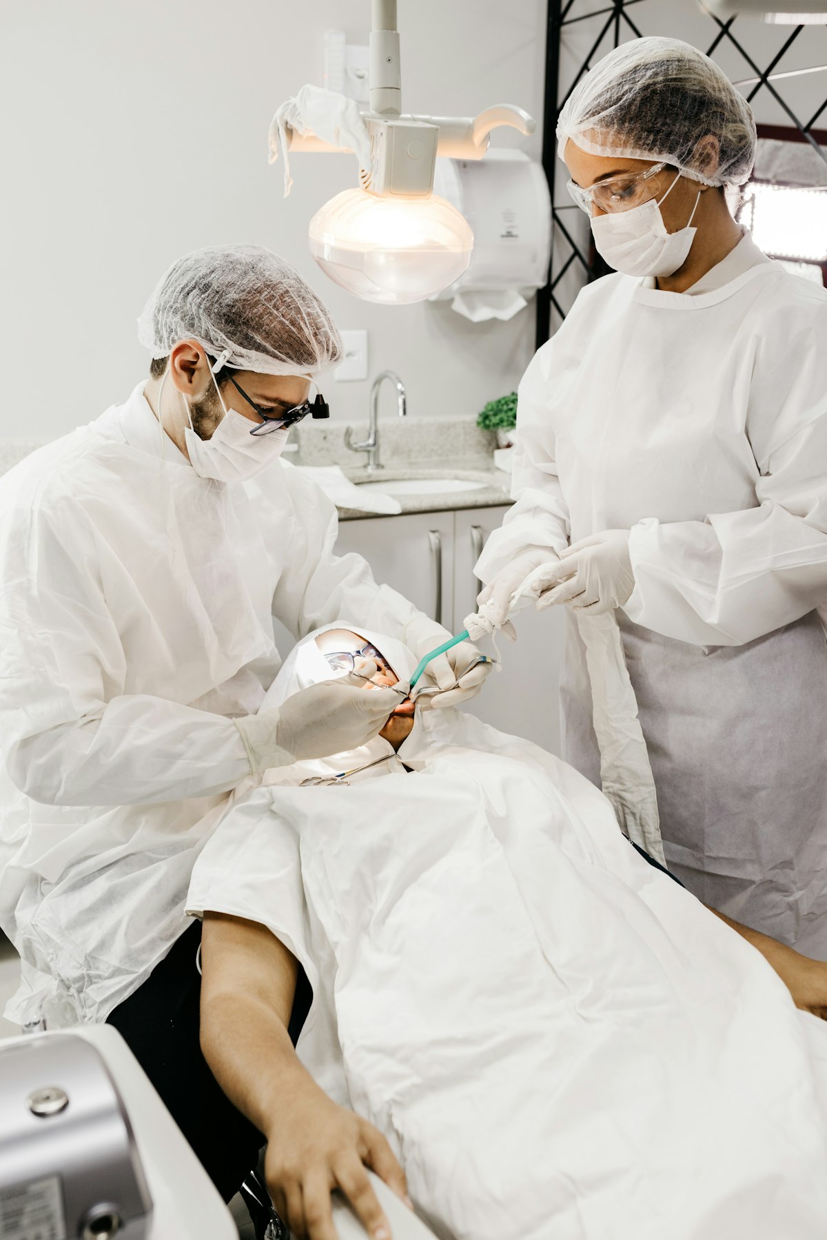
The Thyroid Collar: Protection That Costs Nothing
The thyroid gland is among the more radiosensitive tissues in the body — particularly in children and adolescents. It sits in the neck, positioned directly in the primary beam path for many dental projections, especially periapical and panoramic films.
A thyroid collar — a flexible lead shield that wraps around the neck — measurably reduces thyroid dose in conventional panoramic dental radiography without compromising diagnostic image quality (PMID 24005060). It adds nothing to procedure time and costs essentially nothing to use. Nevertheless, surveys in multiple countries have found that thyroid collars are routinely omitted in dental practice, with usage rates as low as 10–30% in some settings.
The American Dental Association and American Academy of Oral and Maxillofacial Radiology recommend protective shielding and careful patient selection for all patients undergoing conventional dental radiography, particularly children and individuals who are or may be pregnant (PMID 41581943).
The lead apron, by contrast, is primarily a psychological reassurance device for most conventional dental X-rays — the scattered radiation reaching the torso during a bitewing is negligible, and modern equipment with rectangular collimation minimizes it further. This does not mean aprons are useless; for abdominal protection during panoramic imaging in pregnant patients, they retain clear value. But if your dentist uses the apron and forgets the thyroid collar, the more important protection is being skipped.
How Often Should X-rays Actually Be Taken?
Radiographic frequency in dental practice should follow risk-stratified guidelines rather than a fixed schedule. The 2012 ADA/FDA guidelines (updated with subsequent evidence) recommend individualized assessment — a philosophy that sounds obvious but represents a meaningful departure from the historical "annual bitewings for everyone" model.
For adults with low caries risk and healthy periodontal status, posterior bitewing radiographs every 24–36 months is appropriate. For high-risk patients — those with a history of frequent cavities, dry mouth, active periodontal disease, or poor oral hygiene — the interval shortens to 6–18 months. Children follow separate protocols based on eruption status and caries risk (PMID 41581943).
The rationale is straightforward: X-rays are a diagnostic tool, not a screening ritual. They are indicated when the clinical benefit — detecting a lesion that would otherwise be missed and progress — outweighs the (small but non-zero) dose burden. For low-risk patients with good oral hygiene and stable dentition, annual radiography adds little diagnostic value while accumulating unnecessary dose over decades.
Risk-based scheduling requires more clinical judgment and communication than fixed intervals. It also means that two patients sitting in adjacent chairs may receive different imaging recommendations on the same day — which can feel inconsistent to patients unfamiliar with the underlying logic. The dentist's job is to explain that difference, not to standardize away from it.
Cumulative Risk: The Lifetime Perspective
No single dental X-ray causes detectable harm. The linear no-threshold (LNT) model — the dominant framework in radiation protection — holds that any dose, however small, carries a proportional cancer risk, though critics note that at very low doses the absolute risk increase is so small as to be statistically indistinguishable from background variation (PMID 25329961).
Some critics of LNT argue that it overestimates risk at low doses and that below certain thresholds there may be no excess risk at all — a position with genuine scientific support, particularly from studies of populations in high natural background radiation areas who show no excess cancer incidence. The regulatory and clinical consensus, however, is to apply LNT conservatively, accepting some degree of overestimation as the price of caution.
Over a lifetime of dental care, a patient receiving age-appropriate radiographic surveillance might accumulate 500–1,500 μSv from dental imaging specifically. For comparison, living in a city at high altitude (Denver, Mexico City, Bogotá) adds approximately 1,000–2,000 μSv per year in additional cosmic ray exposure relative to sea level. The cumulative dental dose across a 70-year adult life is roughly equivalent to one or two years of altitude-related background differential.
A 2019 systematic review and meta-analysis of the epidemiological evidence (PMID 31502516) found that multiple or repeated dental X-ray exposures may be associated with a small increase in thyroid cancer risk — but the underlying studies were retrospective and subject to recall bias, and the authors stress that thyroid exposure has fallen dramatically with the adoption of thyroid shields and modern low-dose equipment. In other words, the residual concern is itself an argument for the shielding and dose-minimization practices described above.
The question of cumulative risk is worth tracking, but not in a way that leads to diagnostic avoidance. Undetected interproximal caries that progresses to pulpitis, periapical abscess, and tooth loss carries its own cascade of consequences — systemic inflammatory load, the cost and invasiveness of root canal therapy or extraction, the compromised chewing function that follows. Refusing necessary radiography to avoid a dose measured in microsieverts is not a trade that benefits the patient.
What to Ask Your Dentist
Informed patients get better care. The questions worth asking at your next appointment are not confrontational — they are the questions any thoughtful clinician should welcome:
Why are these X-rays being recommended now? A good answer connects the imaging to a specific clinical question: checking for proximal decay not visible on exam, evaluating bone levels in periodontal disease, monitoring a previously identified lesion. "It's been a year" is not a clinical justification.
What type of equipment are you using? Digital detectors require significantly lower exposure than older film-based systems — often 50–80% less. Rectangular collimation versus round collimation reduces dose by approximately 60%. These are not trivial differences.
Will you use a thyroid collar? The answer should be yes, and the collar should go on before the tube is positioned, not as an afterthought.
Is CBCT necessary, or would conventional imaging answer the question? This is particularly relevant before implant placement or orthodontic records.
At QDRO, the underlying belief is that better oral health comes from understanding — not from anxiety, and not from false reassurance. Dental radiography, used appropriately and with proper shielding, is one of the most cost-effective diagnostic tools in preventive dentistry. The numbers support that confidence. Now you have the numbers.
Sources:
- PMID 18762634 — Ludlow JB et al., Journal of the American Dental Association, 2008 — Patient risk related to common dental radiographic examinations under the 2007 ICRP recommendations; effect of collimation
- UNSCEAR 2008 Report — Sources and Effects of Ionizing Radiation — global background radiation averages and effective dose methodology
- PMID 21196094 — Pauwels R et al., European Journal of Radiology, 2012 — Effective dose range for dental cone beam CT scanners (SEDENTEXCT, 14 devices)
- PMID 25270063 — Horner K et al., Dentomaxillofacial Radiology, 2015 — Guidelines for clinical use of CBCT, summarizing the SEDENTEXCT project
- PMID 41581943 — Benavides E et al., Oral Surgery, Oral Medicine, Oral Pathology and Oral Radiology, 2026 — ADA/AAOMR patient selection recommendations for dental radiography and CBCT
- PMID 24005060 — Han GS et al., Dentomaxillofacial Radiology, 2013 — Shielding effect of the thyroid collar for digital panoramic radiography
- PMID 25329961 — Calabrese EJ et al., Radiation Research, 2014 — Critical review of the BEIR VII report and the linear no-threshold (LNT) hypothesis
- PMID 31502516 — Memon A et al., Thyroid, 2019 — Dental X-rays and the risk of thyroid cancer and meningioma: a systematic review and meta-analysis